What Is Syntonic Phototherapy?

Dr. Russel Lazarus, December 20, 2020

absract_colorful_female-1024x682-1

Syntonic phototherapy is a simple, non-invasive light therapy treatment that has been around for more than 70 years.

Syntonic phototherapy uses specific light colors, frequencies and wavelengths to improve the body’s regulatory centers in the brain.

When light enters the eye, it travels through the brain to the pineal gland and the hypothalamus— the areas of the brain responsible for chemical and hormone balance.

According to studies, applying light to these areas can stimulate the brain and help to restore the balance within the body’s nervous system, thereby improving vision problems, such as lazy eye (amblyopia), eye turns (strabismus)convergence insufficiency, and focusing difficulties.

This unique light therapy has also proven effective for the treatment of chronic headaches, stress, learning disorders, trauma, brain injuries and concussions.

If you suspect you have an eye condition, contact an eye doctor near you, who can diagnose and treat the condition.

SEE RELATED: Sleep Apnea, Concussion and Vision

Find a Vision Therapy Eye Doctor Near You

Search near me

How does syntonic phototherapy work?

The colored light used in syntonic phototherapy stimulates the visual system and alters the biochemistry of the brain to improve the balance between the sympathetic and parasympathetic nervous systems.

Colored filter goggles are placed on the eyes for the duration of the light therapy treatment— usually up to 10 minutes. The filter color applied to the goggles is determined based on the presenting visual problem.

Colors such as blue and violet are used to stimulate the parasympathetic nervous system (PNS) which is responsible for controlling our homeostasis and leads to pupil constriction.

Colors on the blue spectrum have been shown to improve focusing abilities for near vision tasks by reducing adrenaline production, anxiety and stress.

Colors such as orange, yellow, and red stimulate the sympathetic nervous system (SNS) responsible for controlling our “fight or flight” response and leads to pupil dilation.

Red and orange are often used to treat lazy eyes, while green and yellow are used to treat esotropia (inward turned eyes).

Can syntonic phototherapy alone treat my vision problem?

In most cases, syntonic phototherapy is prescribed together with vision therapy, specialty lenses and/or prisms to:

  • Improve visual acuity and contrast
  • Increase visual attention
  • Reduce eye strain
  • Decrease light sensitivity

LEARN MORE:  Guide to Neuro-Optometry

To learn more about syntonic phototherapy and determine if you can benefit from this type of treatment, schedule a functional vision evaluation with an eye doctor today.

Posted in Uncategorized | Leave a comment

Foreign Body Removal Start to Finish -these patients are in need of help—quickly. Here’s how to prepare your office.

By Cecelia Koetting, OD

Located in a city with a large shipyard, our office has its fair share of foreign body removal patients. Although incidence and severity are lower with the use of proper protective eye wear, accidents still happen. According to the National Institute for Occupational Safety and Health (NIOSH), each day approximately 2,000 American workers have a job-related eye injury requiring medical treatment.1 NIOSH estimates that 90% of these injuries could be prevented or less severe if the correct eye protection was used.1

It is important for both you and your office staff to feel comfortable handling these emergency patients who require foreign body removal—from start to finish. This article discusses the step-by-step process of triaging, scheduling, examining, treating and following patients with a foreign body.

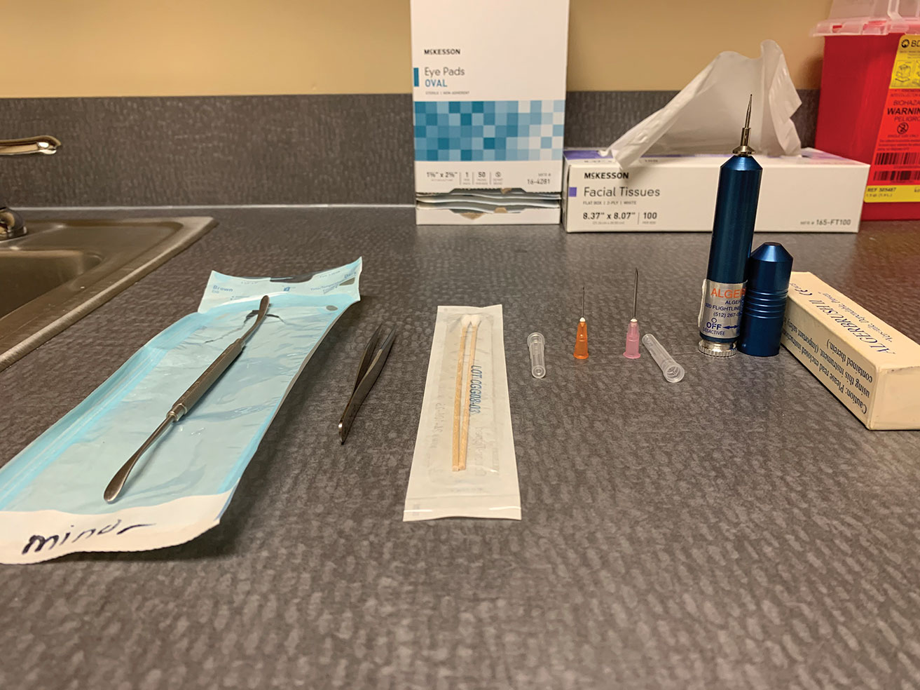
The foreign body removal toolkit includes a spud, jeweler’s forceps, cotton swabs, small-gauge needles and an Alger brush.
Fig. 1. The foreign body removal toolkit includes a spud, jeweler’s forceps, cotton swabs, small-gauge needles and an Alger brush. Click image to enlarge.

Office Prep

Most optometrists have all the tools they need to care for patients with corneal foreign bodies. The entrance tests are common practice for other conditions and include extraocular motilities, pupillary testing and confrontation visual fields. Abnormalities in these could be further indicators of orbital/globe penetration or full penetrating foreign bodies. 

Other standard tests for suspected foreign body include documenting the entering visual acuities prior to any procedure and noting any pre-existing issues of amblyopia or decreased vision.  

For all initial testing, train technicians to handle foreign body removal patients with “no touch” to avoid further irritating the eye or the cornea. Ocular pressures can be checked after the slit lamp exam has been performed, ensuring it is safe. If the foreign body is embedded within the cornea centrally or there is concern for an open globe, it may be best to avoid checking the pressure.

During the slit lamp exam, a topical anesthetic will keep your patient more comfortable, while sodium fluorescein and a cobalt blue filter will help you examine the eye and detect any foreign bodies, wound leakage or a full penetrating injury. If the patient is struggling to keep their lids open, a lid retractor or lid speculum may be necessary.

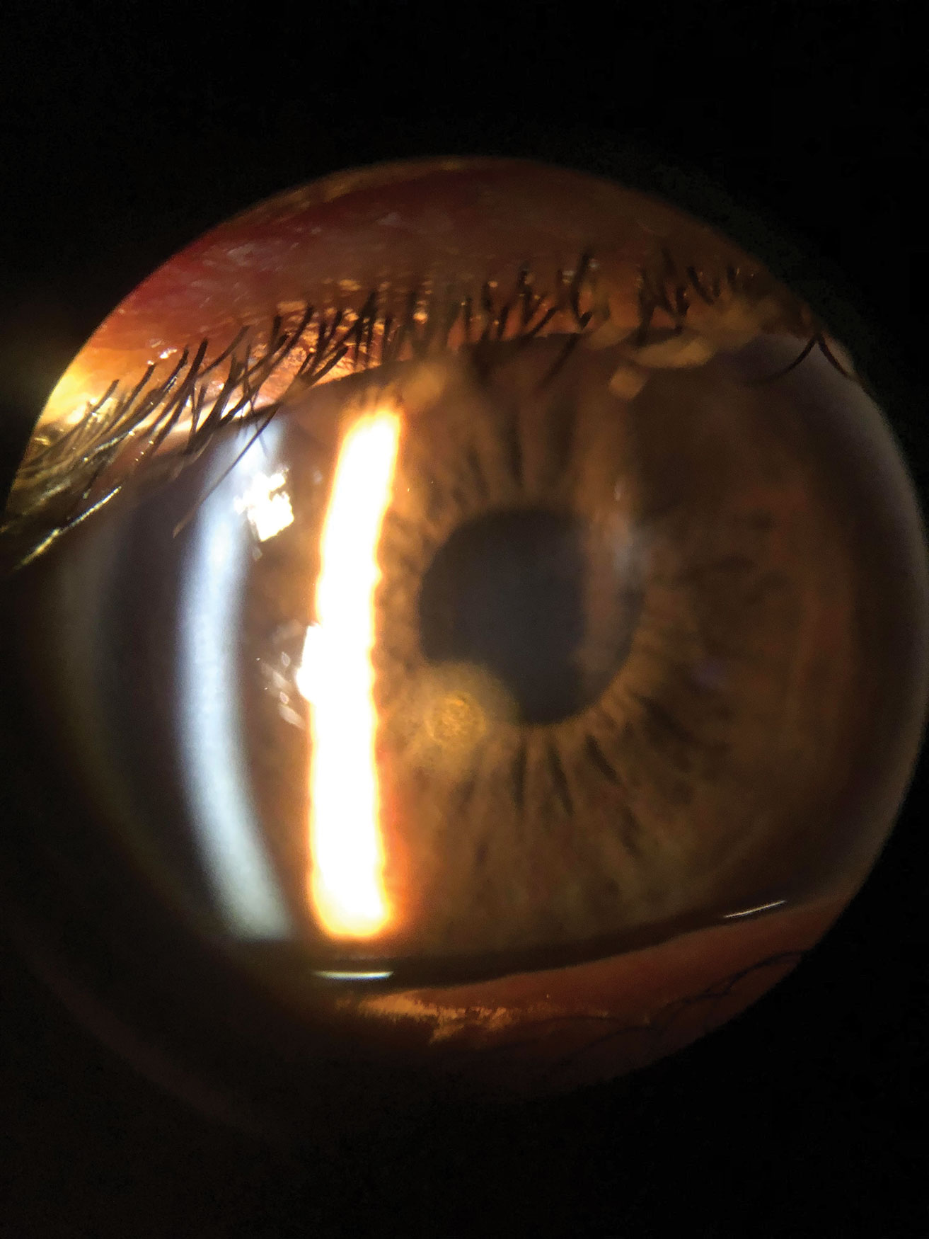
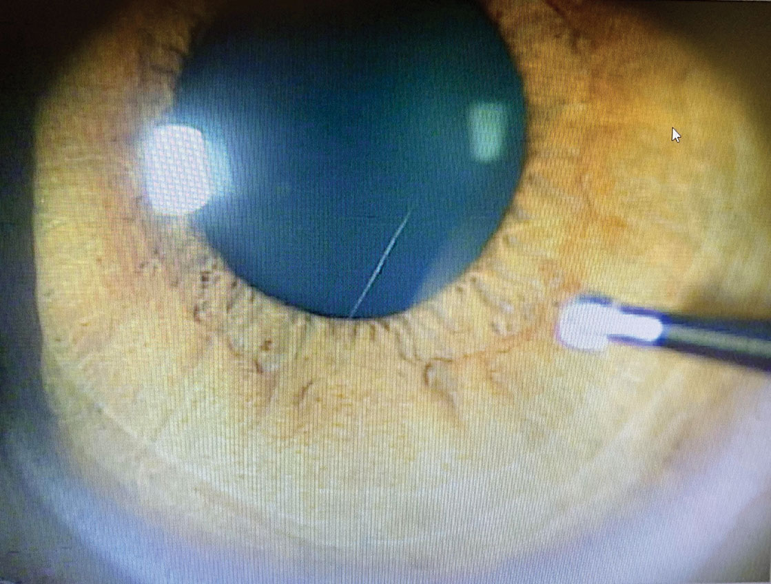
This is a patient’s cornea immediately after removal of a metallic foreign body but prior to rust ring removal. The object was superficial enough that it was removed with a sterile cotton swab after anesthetizing the patient’s eye.
Fig. 2. This is a patient’s cornea immediately after removal of a metallic foreign body but prior to rust ring removal. The object was superficial enough that it was removed with a sterile cotton swab after anesthetizing the patient’s eye. Click image to enlarge.

Clinicians should stock a few different handheld tools in the office to remove various foreign bodies (Figure 1): 

  • Superficial or loosely embedded cornea and conjunctival foreign bodies – The simplest method to remove loose pieces of material may be using only irrigation with sterile saline solution. If that isn’t sufficient, a sterile cotton swab, spatula, spud or a small 25-gauge needle are all useful options. A magnetic spud is quite useful for metallic foreign bodies because you can often remove the object—and any residual metallic flakes—with little to no damage. 
  • Deeper within the corneal stroma – A spatula, spud or a 25-gauge needle are also good options here; while a spud or spatula reduces the risk of perforation, a needle often causes less damage to surrounding tissue. 
  • Protruding corneal and conjunctival foreign bodies – While any of the other methods mentioned may also be used, jeweler’s forceps is an excellent option, as it may allow you to grasp the edge of the object and remove it.
  • Corneal rust ring – This will require an Alger brush to remove.

In addition, stock a topical antibiotic to apply before and after any procedure to ward off infection.

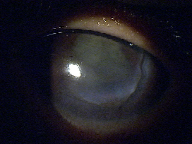
This 13-year-old boy was hit in the eye with his pencil in school, leaving a laceration from 2 o’clock to 9 o’clock. It was pressure patched and seen by cornea specialist, who glued the cornea until the patient could get into the surgical suite, where the surgeon placed 20 sutures to close up the wound.
Fig. 3. This 13-year-old boy was hit in the eye with his pencil in school, leaving a laceration from 2 o’clock to 9 o’clock. It was pressure patched and seen by cornea specialist, who glued the cornea until the patient could get into the surgical suite, where the surgeon placed 20 sutures to close up the wound. Click image to enlarge.

Initial Encounter

When a patient calls or walks in complaining of a foreign body sensation, your front desk staff must understand what to ask the patient to triage and make sure they are seen or referred promptly. Front office and call center staff should begin the patient encounter, whether in person or on the phone, with the tried-and-true five Ws: who, what, where, when and why. These help to ensure patients who need urgent care get it, and those who don’t can be scheduled accordingly. 

For example, a patient who calls saying something got in their eye while doing yardwork yesterday requires different treatment than a patient who had the same thing happen to them a month ago. The latter situation is far less urgent and is less likely a true foreign body than the patient who sustained an injury the day before. 

Patients who mention additional injuries related to the incident may require further testing or treatment. Head trauma requiring imaging or sutures should be sent to the emergency room to address those injuries first. However, keep in mind that MRI is contraindicated for patients with a suspected metallic foreign body. With their serious injuries cared for, the patient can be seen back regarding the ocular foreign body if it wasn’t treated concurrently in the emergency room.

The five Ws can also help to correctly identify patients who may have had a work-related injury associated with a worker’s compensation case. These situations often require extra paperwork to document the details. During documentation, also note if the patient was wearing appropriate personal protective eyewear.

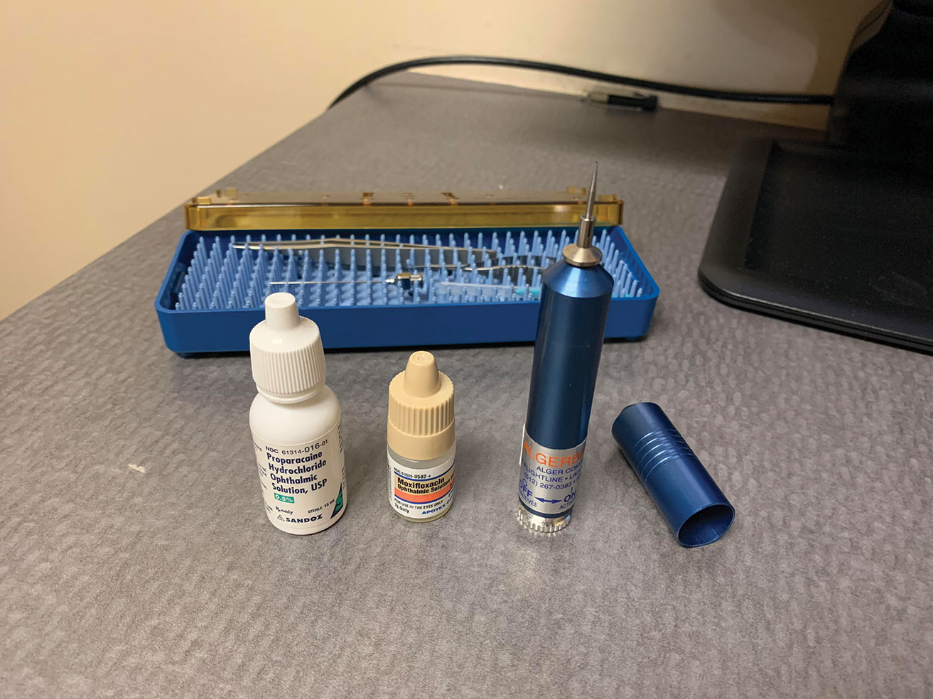
Here is what you will typically need when removing a corneal rust ring with an Alger brush: start with topical proparacaine along with a broad-spectrum topical antibiotic. After the procedure is complete, instill a second drop of antibiotic into the patient’s eye.
Fig. 4. Here is what you will typically need when removing a corneal rust ring with an Alger brush: start with topical proparacaine along with a broad-spectrum topical antibiotic. After the procedure is complete, instill a second drop of antibiotic into the patient’s eye. Click image to enlarge.

In the Chair

Once you are sitting down with the patient, have a detailed discussion with them to understand what exactly is in their eye and how it got there. Depending on the type of foreign body and the timeline, there may be a concern for secondary infections. Patients with suspected vegetative matter warrant higher concern for the development of a fungal corneal ulcer, while a metallic object could cause a rust ring or, more rarely, introduce infectious material leading to bacterial keratitis. 

We also know that the longer the foreign body is in the eye and there is an open abrasion, the higher the risk of developing bacterial keratitis that could lead to an ulcer or even an anterior chamber reaction.2 A study of patients with corneal abrasions due to ocular foreign bodies found that no patients who were seen in clinic within 18 hours of the ocular injury and started on prophylactic antibiotic ointment developed an ulcer.2 However, of those who presented 19 to 24 hours after injury, 3.7% developed an ulcer and 28.6% of those seen between 24 and 48 hours after developed an ulcer.2 

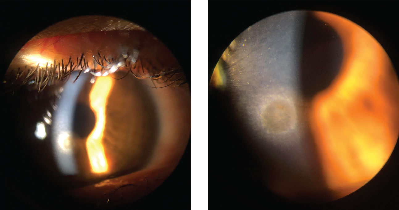
A thorough slit lamp exam will help you determine if the foreign body is still there, its location, depth within the ocular tissue and if it has fully penetrated the cornea (Figure 2). If available, anterior segment OCT can be useful in identifying the depth of the foreign body.3 Patients with a foreign body or abrasion are typically light sensitive and in pain. Instilling a drop of ocular topical anesthetic will help you perform the slit lamp exam. 

Make sure to stain the patient’s eye with sodium fluorescein and use a cobalt blue filter to check for multiple vertical lines on the cornea (tracking patterns) or conjunctival abrasions that may indicate trapped foreign material under or within the lid. Also evert the patient’s lid to visualize all fornicies and ensure no trapped material is causing further corneal damage or later lodge itself within the cornea. 

If the injury occurred from a high-velocity impact or while grinding, it is important to dilate to look for signs that the foreign body has fully penetrated the cornea or globe. Conjunctival penetration is easier to see because there will typically be an area of injection and chemosis surrounding the entrance point. When a conjunctival foreign body or full penetration of the cornea or globe is suspected, check the patient for a positive Seidel’s sign with sodium fluorescein and cobalt blue light filter. Another indication of full penetration with a wound leak would be decreased intraocular pressure or a shallow anterior chamber. 

When looking at the entrance wound on the cornea, check for tracks or disrupted tissue through the cornea stroma or endothelium. An object that has penetrated the ocular lens typically leaves a mark on the anterior portion of the lens, possibly damaging the iris—a wound that can be viewed with iris transillumination. 

Dilation aids in seeing these lenticular marks and helps us visualize the back of the eye to look for the retained object. If you suspect a penetrating intraocular foreign body but cannot directly visualize it, consider sending the patient for orbital radiographs, B-scan or computed tomography to identify and pinpoint the object.4 However, this is not mandatory and may not be a practical use of resources in all cases.

When removing the rust ring from the cornea with the Alger brush, make sure to approach tangentially. This allows for better control of the instrument and pressure applied to the cornea.
Fig. 5. When removing the rust ring from the cornea with the Alger brush, make sure to approach tangentially. This allows for better control of the instrument and pressure applied to the cornea. Click image to enlarge.

Removal Step-by-step

While most of us learned how to remove a foreign body during our education, its rarely an everyday occurrence. Many regional and national conferences provide workshops to help those who feel the need for new or additional training. Before performing any procedure, consult your state law and scope of practice to make sure that you are practicing within your guidelines. 

Before you begin, discuss the steps of the procedure with the patients to reduce their anxiety. In addition, obtain written consent or document in the chart that the patient verbally consented to the treatment plan. If a patient has poor cooperation or is struggling to keep their eyelids open, a lid speculum can help. Have the patient fixate on a target with the opposite eye to help the eye still. Instill a topical anesthetic and a broad-spectrum topical antibiotic.

The location and type of foreign body will often determine how you remove them. Most of us have a preferred method and instrument that we feel comfortable using to remove foreign bodies. 

Superficial or loosely embedded corneal foreign bodies are often easily removed with a sterile cotton swab, spud or a small-gauge needle. 

If the foreign body is deeper within the corneal stroma, use a spud or 25-gauge needle. Using the patients’ forehead for stability, hold the needle head tangentially to the cornea with the beveled tip up, away from the cornea. Place the tip underneath the anterior projection of the foreign body and carefully tease it out. Once dislodged from the stroma, the object can be removed from the surface with a cotton swab or forceps. 

Depending on the practitioner’s comfort level, they should consider referring patients to a cornea specialist. In cases where there is full globe or corneal penetration or a concern for vision-threatening scarring, it is advantageous to consult with a cornea specialist regarding any further treatment or surgical procedures to reduce scarring (i.e., lamellar keratectomy).

Depending on the angle and size of the foreign body, jeweler’s forceps are helpful in cases where the object is protruding from the cornea where an edge is easily accessible or when the foreign body is small such as a splinter or fiberglass.

This patient is being seen at follow-up after a metallic foreign body was removed. While the epithelium has fully healed, there is a remaining rust ring.
Figs. 6 and 7. This patient is being seen at follow-up after a metallic foreign body was removed. While the epithelium has fully healed, there is a remaining rust ring. Click image to enlarge.

If, during the slit lamp and dilation or with anterior segment OCT, you determine the foreign body is full penetrating, consult a surgeon for treatment and removal. If the patient has a positive Seidel’s sign or suspected open laceration, give them a drop of topical antibiotic, pressure patch them to reduce the flow of the leaking aqueous and refer immediately (Figure 3). 

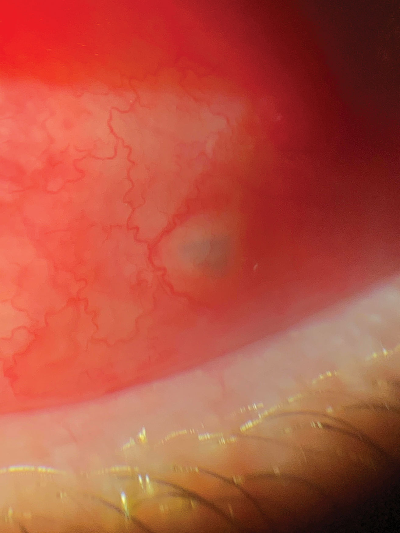
Metallic foreign bodies containing iron may leave behind a rust ring in as little as three to four hours.5 In the event that a rust ring forms, removal is performed using an Alger brush (Figure 4). Use a clean sterile tip and approach the area tangentially with the brush to better control the tool (Figure 5). Any remaining rust ring could cause inflammation and slow or even prevent healing of the corneal epithelial defect. When the rust is deep within the stroma, the patient may require a second treatment with the Alger brush during the follow-up visit to remove the entire rust ring (Figures 6 and 7). However, it is better to remove the entire ring on the initial visit, whenever possible, to reduce the necessity of retreatment.  

When the foreign body seats itself within the conjunctiva, clinicians may be able to remove it in-office without surgery (Figure 8). The entering wound, if shallow and recent, sometimes functions as an access point for removal with jeweler’s forceps. If it has been more than 48 hours or the material is seated too deeply, the removal may require incision and possible sutures and, in some cases, may warrant a referral. 

Bacterial cultures are not routinely performed on patients with corneal foreign bodies without a stromal infiltrate. One study found that only nine of 63 tested corneal foreign bodies were positive for bacteria.6 

After any foreign body removal, scan the patient’s eye to ensure no other possible foreign bodies are present and document the extent of the patient’s treatment area. 

Most importantly, educate the patient prior to leaving the office regarding proper protective eye wear to help decrease the likelihood of re-occurrence.  Discuss not only what is considered proper protective eye wear but also when they should be wearing it (e.g., yard work, working on cars, grinding, cutting).

This patient presented to the office within eight hours of being hit in the eye with a tree branch. A piece of bark had lodged itself under the conjunctiva. The foreign body was removed with forceps through the entry point with no sutures needed.
Fig. 8. This patient presented to the office within eight hours of being hit in the eye with a tree branch. A piece of bark had lodged itself under the conjunctiva. The foreign body was removed with forceps through the entry point with no sutures needed. Click image to enlarge.

Homework

Patients should be started on a topical antibiotic immediately following the removal of the foreign body. Typically, a broad-spectrum topical antibiotic drop such as a flouroquinolone QID is appropriate, given the patient has no allergies. In the case of conjunctival involvement or large abrasions, the patient may be more comfortable using an ophthalmic antibiotic ointment such as tobramycin or Polytrim (polymyxin B/trimethoprim, Allergan) BID to QID. If vegetative matter involvement is suspected and a fungal ulcer is present, the patient should be started on topical and oral anti-fungal medication. 

Pain management for these patients can vary depending on situation and the patient. The use of a therapeutic bandage contact lens, while not necessary, can provide sufficient relief but should be avoided if there is any formation of secondary ulcer at the time of exam. Some patients find comfort from pressure patching the eye, although this can be cumbersome with the requirement of frequent ocular drops. To avoid this, it is preferable to use ophthalmic antibiotic ointment, instilled prior to pressure patching. Topical nonsteroidal anti-inflammatory agents can aid in pain management, as can atropine. 

Oral analgesics and anti-inflammatory medications such as Tylenol and ibuprofen may be added as needed. The use of narcotics can be considered on a case-by-case basis in appropriate patients but is often unnecessary. 

Patients with larger lesions and those with a bandage contact lens or pressure patch should have a follow up in 24 hours. Smaller or peripheral lesions can have an extended follow up of up to a week for non-complicated cases if no ulcer or infection is present. When an ulcer has formed or infection is present, the patient should be seen the next day to monitor for improvement. 

Amniotic membranes or amniotic drops can also be considered for patients with deep central foreign bodies.7 If the patient’s injury was due to vegetative matter, it is important to watch for possible infiltrate and ulcer development prior to starting any steroid, as this will worsen a fungal infection. 

Knowing how to help established or new patients with an urgent foreign body situation is essential for your community and your practice. The need for foreign body removal is a common and frequent complaint. As optometrists, we should feel comfortable in treating, or in the very least, triaging these patients. Once you establish yourself as knowledgeable and competent, you will enjoy increased personal fulfillment—and referrals.

Dr. Koetting is the referral optometric care and externship program coordinator at Virginia Eye Consultants in Norfolk, VA. She is a fellow of the American Academy of Optometry and a trustee of the Virginia Optometric Association.

Posted in Uncategorized | Leave a comment

Children’s myopia risk linked to smartphone use, study says

JUNE 15, 2020Researchers found an association between myopia and prolonged smartphone use, begging the question: What does the future hold for millions of kids growing up in the digital age?

Device use Myopia in children

Myopic children use twice as much smartphone data each day as their non-myopic peers, a recent study found, leading researchers to ponder whether iGen’s unparalleled connectivity from such a young age exacerbates myopia risk.
 
Published online in the journal Clinical and Experimental Optometry, the study found myopic refractive error was associated with increased smartphone data usage as normal-vision children consumed an average of 614 MB data daily and spent nearly 4 ½ hours on the phone daily, whereas myopes consumed an average of 1,131 MB data daily and spent nearly 5 hours on the phone daily. The findings add to a growing body of evidence detailing the effects of increased device use on refractive error and raise concerns over the next generation of Americans-iGen or Gen Z-growing up with a preponderance of digital devices.  

“Based on the results from both studies, the risk for myopia development and progression may be higher in this pandemic situation.”

Although more than 9 in 10 (93%) millennials own smartphones, leading all other American generations in adoption and use of technology, the generation now spanning ages 24 to 39 doesn’t hold the distinction of “digital natives.” That title belongs to Gen Z, those born after 1996, who have no memory of a world before smartphones. In fact, 95% of American teens own or have access to a smartphone and nearly half say they’re online on a near-constant basis.  

As digital device use has skyrocketed in recent years, so, too, has myopia prevalence and age of onset. In just four decades, U.S. myopia prevalence has gone from 25% of Americans to about 42%, while a 2018 study of 60,800 children in southern California found 59% of 17- to 19-year-olds were myopic. And, if that wasn’t dramatic enough, myopia prevalence globally could reach 52% in the next three decades. Hence, the impetus behind this international study originating from the University of Dublin.  

Smartphone usage associated with myopia  

Per the study, researchers posed a questionnaire on smartphone usage to 418 Irish students enrolled in primary (K-6), secondary (7-12) and tertiary (university) level schooling, then compared their refractive status. Students’ smartphone data usage was recorded over an extended period, as were the top three applications consuming most data and a self-report of estimated time spent per day on the phone.  

Students used an average of 873 ± 1,038 MB (mean ± standard deviation) of data daily and about four hours of time spent on the phone each day, mostly spent using social media apps (Snapchat, Instagram and Facebook). Researchers found that myopes not only used more data (1,131 ± 1,748 MB data/daily compared to 614 ± 902 MB data/daily) and spent longer on their phones than non-myopes but also were more likely to believe screen use could affect their eyes.  

Interestingly, 84% of myopes believed digital technology could adversely affect their eyes as compared to 68% of non-myopes with both groups citing dry eyes (67%) and eyestrain (29%). A similar number of myopes and non-myopes believed digital screen use could cause myopia (31% and 25%, respectively).  
“The lifestyle habits of children and teenagers today have undeniably changed with advancements in technology and while the prevalence of myopia has been increasing for decades, the increased level of near visual stimulation from smartphones may pose an additional independent risk for myopia,” the authors note.  

“Smartphones differ from traditional reading in various aspects such as wave-length, distance from the eye, size, contrast, resolution, temporal properties and spectral composition, all of which merit investigation. Aside from this, children and adolescents now spend more (time) than ever using a smartphone that demands proximal attention, which may compete with other more protective activities such as time outdoors.”  

Corroborating evidence

This latest study isn’t the only one to find an association between myopia and smartphone use. In fact, a 2019 PLoS One study also concluded an association in smartphone and computer usage with greater refractive error yet did not find an association when it came to television viewing or after-school study. Additionally, the PLoS One study reported a statistically significant association between outdoor time and reduced myopia, but only for the noonday timeframe when illumination is greatest.  

Yi Pang, M.D., O.D., Ph.D., associate dean for research at Illinois College of Optometry, says there is a trend that more children are spending more time on digital devices, and research has shown that longer time on near work and/or shorter working distance can increase myopia progression. This should be a red flag as schoolchildren only recently concluded months of distance learning on handheld devices or computers due to pandemic lockdowns.

 “With the COVID-19 pandemic, the time on electronic devices dramatically increased in children because of remote learning and fewer opportunities for kids to do other activities,” Dr. Pang says. “Based on the results from both studies, the risk for myopia development and progression may be higher in this pandemic situation.”  
However, Dr. Pang notes, both studies only found an association with myopia and not a causation, which should be grounds for future studies.  
Dr. Pang adds: “Eye care practitioners should be aware of this issue and be ready to address parents’ and patients’ concerns.”  

For more patient resources, access AOA’s digital eyestrain and blue light information, or visit AOA Marketplace for patient education products available on blue light.

Posted in Uncategorized | Leave a comment

Seven Myths About Sunglasses That Could Damage Your Vision

Nation’s ophthalmologists debunk common myths, offer tips to promote smart eye health choices

SAN FRANCISCO – Did you know that too much sun on unprotected eyes increases the risk of eye disease? The good news is that prevention is simple: Wear sunglasses that block 99 to 100 percent of UVA and UVB radiation. But there are a lot of common myths about sunglasses and eye health. That’s why the American Academy of Ophthalmology is working to debunk the myths and offer tips to promote smart eye health choices.

  • Myth #1: Sunglasses are only for sunny days
    False. UV light gets through clouds and haze. Regardless of the season, sun exposure can increase the risk of developing cataractsgrowths on the eye and cancer. Sun reflecting off ice and snow can also lead to photokeratitis, a painful eye condition sometimes referred to as snow blindness.
  • Myth #2: Babies and children don’t need to wear sunglasses
    False. Children are just as susceptible to the sun’s harmful rays as adults. Start them on healthy habits early.
  • Myth #3: Cheap sunglasses don’t offer protection
    False. Sunglasses don’t have to cost a lot of money to provide adequate eye protection. Less expensive pairs marked as 100 percent UV-blocking can be just as effective as more expensive options. Labels can sometimes be confusing. Some indicate sunglasses offer 100 percent protection from UVA/UVB radiation, others offer 100 percent UV 400 protection. Both block 100 percent of the sun’s harmful radiation.
  • Myth #4: Size doesn’t matter
    False. The more coverage sunglasses provide, the less sun damage inflicted on the eyes. Consider oversized sunglasses or wraparound-style glasses, which can help cut down on UV light entering the eye from the side. Wraparound glasses may also protect against dry eye due to the wind.
  • Myth #5: Polarized lenses offer more protection from the sun
    False. Polarized lenses do not block more radiation; however, they can cut down on glare reflecting off cars, water or pavement, which can make activities such as driving or boating more enjoyable.
  • Myth #6: Darker lenses offer more protection from the sun
    False. The darkness of the lens isn’t indicative of protection. Make sure the label on the lens says 100% UV-blocking.
  • Myth #7: Tinted lenses offer more protection from the sun
    False. Amber, green and grey lenses do not block more of the sun’s harmful rays. However, tinted lenses do offer increased contrast for those who play sports, making a softball or a golf ball easier to see.

“Your eyes need protection from the sun’s damaging ultraviolet rays just like your skin,” said Dianna Seldomridge, MD, clinical spokesperson for the American Academy of Ophthalmology. “Sunglasses are your prescription for eye health. They are effective, inexpensive, and provide protection year-round.”

For more information about eye health, visit the Academy’s EyeSmart website.


About the American Academy of Ophthalmology
The American Academy of Ophthalmology is the world’s largest association of eye physicians and surgeons. A global community of 32,000 medical doctors, we protect sight and empower lives by setting the standards for ophthalmic education and advocating for our patients and the public. We innovate to advance our profession and to ensure the delivery of the highest-quality eye care. Our EyeSmart® program provides the public with the most trusted information about eye health. For more information, visit aao.org.

Posted in Uncategorized | Leave a comment

Therapy for common vision disorder fails to improve children’s reading test scores

Results from a clinical trial funded by the National Eye Institute (NEI) show that while vision therapy can successfully treat convergence insufficiency (CI) in children, it fails to improve their reading test scores. Investigators from the Convergence Insufficiency Treatment Trial – Attention and Reading Trial (CITT-ART) published the results online today in Optometry and Vision Science. NEI is part of the National Institutes of Health.

CI is a common childhood vision disorder in which the eyes are unable to work together when looking at nearby objects, and the condition can make tasks like reading difficult. Eye care providers who treat CI have assumed that successful vergence/accommodative therapy, often called vision therapy, would lead to better reading fluency and comprehension.

While in-office vision therapy can improve visual function for children with CI, this trial indicates that clinicians should not suggest that it will lead to increased reading performance.”

Lead study author Mitchell Scheiman, O.D., Ph.D., Salus University, Elkins Park, Pennsylvania

When reading or performing other close work, the eyes must turn inward to converge. For children with CI, the eyes have difficulty converging accurately, which can lead to blur or double vision, causing symptoms like discomfort and difficulty maintaining concentration when reading. Results from the earlier Convergence Insufficiency Treatment Trial (CITT), published in Ophthalmology in 2008, showed office-based vision therapy to be the most effective treatment for improving convergence and ameliorating symptoms, and that treatment effects were long-lasting. This new clinical trial, CITT-ART, was designed to determine whether treating symptomatic CI would improve reading skills.

The study enrolled 310 children with symptomatic CI, ages 9 to 14 years, and randomly assigned them to two groups. About two-thirds (206) received in-office vision therapy for 16 weeks, and the remainder (104) received in-office placebo therapy for 16 weeks. The in-office vision therapy was designed to improve accommodation (focusing) and vergence (eye teaming, meaning the ability of the eyes to work together), while the placebo therapy procedures were designed to have no effect on CI.

The children’s reading performance and clinical signs of CI were measured when the study began and again after completing 16 weeks of therapy. Reading comprehension was assessed using the Wechsler Individual Achievement Test- Version 3 (WIAT-III). Additional assessments included word reading and pseudoword decoding (sounding out fake words), as well as oral and silent reading fluency. Clinical signs included measures of how well the eyes work together to converge on near objects. Symptoms were measured using the Convergence Insufficiency Symptom Survey.

While all children showed improved reading comprehension on the WIAT after 16 weeks, there was statistically no difference between average reading improvement for the CI treatment group (3.68 points) and placebo group (3.80 points). Nor was in-office vision therapy better than the placebo therapy on other standardized reading tests. Meanwhile, 75-80% of children in the vision therapy group had shown significant improvement and fell into the normal range for clinical signs, compared with approximately 30% in the placebo group. These results are similar to the original CITT study. However, the latest findings show no significant difference in symptoms, with 62% of children in the vision therapy group versus 58% of children in the placebo therapy group reporting significant symptom improvement. These findings suggest that clinical measures, rather than self-reported symptoms, are critical for assessing CI severity and improvement in children.

Posted in Uncategorized | Leave a comment

Important coronavirus updates for ophthalmologists

APR 26, 202
CDC, WHO
Comprehensive Ophthalmology

Returning to Ophthalmology Practice
A message from Academy CEO, David W. Parke II, MD, on reopening ophthalmology care.

List of Urgent and Emergent Ophthalmic Procedures
In collaboration with major subspecialty societies, the Academy has collated a list of urgent and emergent procedures generally performed in operating rooms at hospitals or ambulatory surgery centers.

For complete coverage of the COVID-19 pandemic, visit the Academy’s resource page Coronavirus and Eye Care.

Updated April 26, 2020, 10 p.m. PT. The following sections have been updated or added in the past 48 hours: Ophthalmology ties

The Academy is sharing important ophthalmology-specific information related to the novel coronavirus, referred to as severe acute respiratory syndrome coronavirus 2 (SARS-CoV-2). The highly contagious virus can cause a severe respiratory disease known as COVID-19.

This page is principally authored by James Chodosh, MD, MPH, with assistance from Gary N. Holland, MD, and Steven Yeh, MD.

What you need to know

  • Several reports suggest that SARS-CoV-2 can cause a mild follicular conjunctivitis otherwise indistinguishable from other viral causes, and possibly be transmitted by aerosol contact with conjunctiva. However, at this point in the COVID-19 pandemic, practically any patient seen by an ophthalmologist could be infected with SARS-CoV-2, regardless of presenting diagnosis, risk factors, indication for visit or geographic location.
  • SARS-CoV-2 is susceptible to the same alcohol- and bleach-based disinfectants that ophthalmologists commonly use to disinfect ophthalmic instruments and office furniture. To prevent SARS-CoV-2 transmission, the same disinfection practices already used to prevent office-based spread of other viral pathogens are recommended before and after every patient encounter.
  • The Academy and federal officials strongly recommend protection for the mouth, nose and eyes when caring for patients potentially infected with SARS-CoV-2.

Background

SARS-CoV-2 is an enveloped, single-stranded RNA virus that causes COVID-19. It is highly transmissible and has a significant fatality rate, especially in the elderly and those with comorbidities such as immune suppression, respiratory disease and diabetes mellitus. A significant number of global fatalities have occurred, and the impact is being felt worldwide.

Patients typically present with respiratory illness, including fever, cough and shortness of breath; diarrhea is common early in infection, and conjunctivitis has also been reported. Other less specific symptoms include headache, eye pain and fatigue. Complications in severe cases include pneumonia, renal failure, cardiomyopathy, stroke and encephalopathy. Symptoms can appear as soon as 2 days or as long as 14 days after exposure. A March 10 study in the Annals of Internal Medicine found that the mean incubation period for SARS-CoV-2 was 5 to 7 days, and patients were typically infectious for several days prior to symptom onset. More than 97% of those who developed symptoms did so within 11.5 days of exposure, findings that further support current 14-day quarantine recommendations.

Transmission

Current understanding about how COVID-19 spreads is based largely on what is known about other similar coronaviruses. The virus is believed to spread primarily via person-to-person through respiratory droplets produced when an infected person coughs or sneezes. It also could be spread if people touch an object or surface with virus present from an infected person, and then touch their mouth, nose or eyes. Viral RNA has also been found in stool samples from infected patients, raising the possibility of transmission through the fecal/oral route.

Existing evidence suggests that SARS-CoV-2 is commonly spread by asymptomatic and presymptomatic transmission. A Feb. 21 report in JAMA details a case of an asymptomatic carrier who possibly infected 5 family members despite having normal chest computed tomography (CT) findings. In an outbreak at a long-term care skilled nursing facility in King County, Washington, 30% of residents tested positive for SARS-CoV-2; half of these were asymptomatic on the day of testing. In another study from China, the transmission rate from those with “undocumented” infections (asymptomatic or symptomatic but not meeting testing criteria) was lower but still significant, with an estimated transmission rate about half that of those with classical symptoms. Because undocumented infections are more common than current testing levels can account for, they may serve as a greater source of transmission than those of symptomatic carriers who have tested positive. The median duration of viral shedding in a study of 191 COVID-19 positive inpatients in China was 20 days; the longest duration observed was 37 days. Li Wenliang, MD, the whistleblower ophthalmologist who sounded the initial alarms on the coronavirus, and later died from the disease, believed he was infected by an asymptomatic glaucoma patient. The sum of these reports points to asymptomatic and presymptomatic transmission as a significant source of spread.

Environmental contamination by SARS-CoV-2 is another cause for concern. In a study published in the New England Journal of Medicine, scientists were able to detect viable SAR-CoV-2 in aerosols up to 3 hours post-aerosolization, although in an experimental setup lacking any ventilation, and not necessarily reflecting how the virus behaves in real-life conditions. The study also found infectious virus could survive up to 24 hours on cardboard, up to 4 hours on copper, and up to 2 to 3 days on plastic and stainless steel. In another study by the CDC of the recent cruise ship outbreaks of COVID-19, SARS-CoV-2 RNA (not necessarily indicating infectious virus) was identified on various surfaces within cabins of passengers who tested positive, for up to 17 days after they disembarked the ship.

In a report from the University of Nebraska, the authors used RT-PCR to test room air, personal articles and environmental surfaces for SARS-CoV-2 in the hospital rooms of COVID-19 patients. They found evidence of viral RNA throughout the rooms—even on windowsills and in ambient air. However, infectious virus could not be cultured from air samplers. These data emphasize the need to use personal protective equipment (PPE) meticulously, per local guidelines, and to remain vigilant in handwashing and disinfecting surfaces and materials possibly contaminated by respiratory secretions from infected patients.

Personal protective equipment (PPE) for ophthalmic use

There is heightened controversy regarding what constitutes appropriate PPE for ophthalmologists performing ophthalmic examinations, particularly around the use of masks and goggles. Reports of ophthalmologist and otolaryngologist deaths in China and Italy, new data about environmental virus contamination and increased awareness of asymptomatic and presymptomatic spread of new infections all favor mouth, nose, and eye protection. However, global shortages of PPE and concerns about mask effectiveness with extended wear and reuse have thus far impacted widespread adoption. Consequently, U.S. hospital guidance has varied from forbidding physicians from wearing masks except in high-risk interactions, presumably for fear of shortages, to mandating all hospital staff and patients wear surgical masks to reduce asymptomatic transmission. In hotspots, some hospitals require all caregivers to wear N-95 masks. The Academy relies on expert guidance from the CDC with regards to indications for extended mask wear and reuse. The use of masks during ophthalmic examinations is a rapidly evolving issue and our editors will update this page as new recommendations emerge.

Treatment and vaccine

Currently, there are no proven agents for prophylaxis or therapy for SARS-CoV-2 infection. Prominent among those under study are chloroquine and hydroxychloroquine, oral agents approved for malaria and autoimmune disorders, respectively, but concerns remain about potential cardiotoxicity. Remdesivir, an investigational antiviral drug, has shown promise in a recently published compassionate use study. Monoclonal antibody IL-6 receptor antagonists are being studied to address the “cytokine storm” seen in some patients with severe COVID-19. There are also efforts to use convalescent sera from COVID-19 survivors as therapy. More information about developments in the treatment of COVID-19 by these agents is available from the CDC.

At this time, there is no vaccine to prevent infection, but on March 5, mRNA coronavirus vaccine trials began enrollment at Kaiser Permanente Washington Health Research Institute in Seattle, and at Emory Children’s Center in Decatur. Both institutions are members of the Infectious Diseases Clinical Research Consortium, a clinical trials network supported by the National Institute of Allergy and Infectious Diseases (NIAID).

Use of chloroquine and hydroxychloroquine

The American Academy of Ophthalmology has no opinion on the use of chloroquine or hydroxychloroquine in COVID-19 patients. However, in a review of published guidelines for the use of these 2 drugs as treatment for COVID-19, a working group* from the Asia-Pacific Vitreo-Retina Society found that proposed doses in many of the ongoing studies worldwide exceeded the maximum daily dose considered safe for long-term therapy (generally <5mg/kg of real weight for hydroxychloroquine) for rheumatic and other chronic diseases (WF Mieler, MD, personal communication, March 25, 2020).

The risk of irreversible maculopathy at these higher doses for short periods of time is unknown. Patients should be informed of the potential for macular toxicity before starting therapy. Furthermore, the need for baseline fundus examination and/or imaging is also unknown in cases with high doses over a relatively short duration. Additional diagnostic testing, such as an ERG, prior to placing a patient on hydroxychloroquine for treatment of COVID-19 is likely unnecessary due to the short treatment duration. Additionally, performing an ERG in this setting carries an unnecessary risk of virus transmission. Until more is learned about the toxicity associated with current regimens, decisions should be made on an individual basis, taking into consideration any pre-existing retinal disease. As in all cases, the Academy urges ophthalmologists to make decisions guided by available scientific evidence.
Ruamviboonsuk P, Lai T, Chang A, Lai C, Mieler W, Lam D

Updated Ophthalmology ties

Several published reports and a recent news article suggest that SARS-CoV-2 can cause conjunctivitis, either as an early sign of infection, or during hospitalization for severe COVID-19 disease. Thus, it is possible that SARS-CoV-2 is transmitted to the conjunctiva by aerosol or through hand to eye contact. There is also evidence for SARS-CoV-2 RNA in tears of COVID-19 patients with conjunctivitis, although infectious virus has not yet been cultured from the conjunctiva of any COVID-19 patient.

  • In a Journal of Medical Virology study of 30 patients hospitalized for COVID-19 in China, 1 had conjunctivitis. That patient—and not the other 29—had SARS-CoV-2 RNA in ocular secretions. This suggests that SARS-CoV-2 can cause conjunctivitis, and that infectious viral particles might be present in tears of COVID-19 patients with conjunctivitis. This observation was confirmed in a recent case report from China and another from Italy of patients with conjunctivitis in the setting of COVID-19.
  • In a larger study published in the New England Journal of Medicine, researchers documented “conjunctival congestion” in 9 of 1,099 patients (0.8%) hospitalized with laboratory-confirmed COVID-19 from 30 hospitals across China. None of the patients were documented to have seen ophthalmologists, and tears were not sampled.
  • In a retrospective case series published March 31 in JAMA Ophthalmology, 12 of 38 “clinically confirmed” hospitalized cases of COVID-19 in Hubei Province, China, had ocular “abnormalities,” characterized most commonly as chemosis and/or secretions. Two patients had a positive conjunctival swab for SARS-CoV-2 RNA, one with signs of conjunctival hyperemia and the other with chemosis and epiphora. This paper was discussed in the New England Journal of Medicine Journal Watch, which stated that “a third [of patients in the JAMA Ophthalmology paper] had conjunctivitis.” We disagree with the latter characterization; chemosis in a critically ill patient most likely represents third-spacing or fluid overload, not conjunctivitis.
  • Two preprint studies posted on MedRxiv also suggest a relatively low likelihood of infectious virus in tears of COVID-19 patients. In a study by Zhang et al. of 72 confirmed COVID-19 patients at Tongji Medical College, 2 patients had conjunctivitis. One of the 2 with conjunctivitis and none of the other 70 patients had SARS-CoV-2 RNA in their tears. In a paper by Zhou et al. of 63 confirmed COVID-19 patients in Wuhan, only 1 had conjunctivitis; that patient had a negative conjunctival swab for SARS-CoV-2 RNA. One other patient’s conjunctival swab was positive and 2 were “probable.”
  • In a case report published in the Annals of Internal Medicine, the first patient in Italy to be diagnosed with COVID-19 also had conjunctivitis in addition to fever and respiratory and gastrointestinal signs. RT-PCR on conjunctival swabs showed SARS-CoV-2 RNA from day 3 of hospitalization until day 21 (1 day after the conjunctivitis resolved), and again at day 27, at which point nasal swabs were negative. Infectious virus was isolated by cell culture from a sample taken on day 3.
  • In a story from CNN, a registered nurse in a nursing home in Washington state with a large COVID-19 outbreak reported that red eye was a common early sign in elderly patients who then became sick with COVID-19.

Unless or until the CNN report is confirmed, existing data suggest that conjunctivitis is an uncommon event as it relates to COVID-19. However, because conjunctivitis is a common condition overall, and patients with conjunctivitis frequently present to eye clinics or emergency departments, it may happen that ophthalmologists are the first providers to evaluate patients possibly infected with COVID-19. Based on the studies above, it is possible that a patient with COVID-19 associated conjunctivitis could have infectious virus in their ocular secretions.

Therefore, protecting your mouthnose (e.g., an N95 mask) and eyes (e.g., goggles or shield) is recommended when caring for patients potentially infected with COVID-19. In addition, slit-lamp breath shields (e.g., here ) are helpful for protecting both health care workers and patients from respiratory illness. Free slit-lamp breath shields are being offered by some manufacturers, including Topcon and Zeiss.

Questions you should ask to identify patients with possible exposure to SARS-CoV-2

  • Does your patient have sore throat, fever, fatigue, loss of smell or respiratory symptoms?
  • Has your patient been in the presence of someone with known COVID-19 in the last 2 to 14 days?

In regions currently managing significant outbreaks of COVID-19, it is safest to assume that any patient could be infected with SARS-CoV-2, and to proceed accordingly. The CDC is urging health care providers who encounter patients meeting these criteria to immediately notify both infection control personnel at your health care facility and your local or state health department.

Outpatient clinics and elective surgery

On March 18, the Academy issued a statement urging all ophthalmologists to immediately cease providing any treatment other than urgent or emergent care. That statement was made based on recommendations from the American College of Surgeons and the CDC. Since then, based on the April 16 guidelines from the federal government, some facilities have begun to prepare for resuming elective surgeries.

Decisions on reopening eye clinics to routine care and resuming elective eye surgery will need to be made in consideration of numerous factors, as recently outlined by the American College of Surgeons and other leading medical organizations. These include but are not limited to evolving city and state restrictions to nonessential services, local/regional new case rates, availability of PPE and access to COVID-19 testing.

Read the full message from Academy CEO, David W. Parke II, MD, on reopening ophthalmology care.

Interim guidelines on resuming elective ophthalmic care

Until there is reliable point-of-care testing, an FDA-approved and highly effective therapy and/or an approved and effective vaccine, practices and clinics should continue to mandate social distancing in waiting rooms, frequent and meticulous disinfection of patient waiting and care areas, and the wearing of face coverings by both patients and caregivers. This means that clinic schedule volumes might need to remain below pre-COVID-19 levels for the foreseeable future. Additional precautions required in operating rooms may lead to longer turnover times between cases, thus impacting the number of surgical cases that can be performed per session. Presuming compliance with state and local regulations, the Academy recommends resumption of normal activities be undertaken gradually, in order to accommodate these and other as yet unforeseen issues.

The Academy also recognizes the potential role of serologic testing for ophthalmologists and other health care providers when considering resumption of elective visits and surgeries, but for now suggests caution in the interpretation of testing results. In response to the pandemic, current FDA policy allows developers to market serologic tests for antibodies to SARS-CoV-2. However, the FDA has not yet undertaken independent reviews of these tests for accuracy and reliability. A false-positive serologic test in an asymptomatic health care worker could leave them unknowingly at risk for COVID-19. Because humoral antibody responses to SARS-CoV-2 typically appear within 1 to 2 weeks of infection, while infected individuals can shed virus for up to 5 weeks from the onset of infection, a person with positive serology after asymptomatic infection might still be shedding virus and therefore remain infectious to patients and coworkers. It is also unknown to what degree a positive IgG response confers resistance to reinfection with SARS-CoV-2, and for how long. Finally, in regions with low prevalence of COVID-19, there is increased likelihood that a positive serologic test result would be an artifact or a testing error rather than reflect prior asymptomatic infection.

Recommended protocols when scheduling or seeing patients

  • If the office setup permits, patients who come to an appointment should be asked prior to entering the waiting room about fever and respiratory illness and whether they or a family member have had contact with another person with confirmed COVID-19 in the past 2 to 14 days. If they answer yes to either question, they should be sent home and told to speak to their primary care physician about testing (see interim guidance below).
  • Keep the waiting room as empty as possible, advise seated patients to remain at least 6 feet from one another. As much as prudent, reduce the visits of the most vulnerable patients.
  • The use of commercially available slit-lamp barriers or breath shields is encouraged, as they may provide a measure of added protection against the virus. These barriers do not, however, prevent contamination of equipment and surfaces on the patient’s side of the barrier, which may then be touched by staff and other patients and lead to transmission. Homemade barriers may be more difficult to sterilize and could be a source of contamination.
  • To further decrease the risk of viral spread, ophthalmologists should inform their patients that they will speak as little as possible during the slit-lamp examination, and request that the patient also refrain from talking.
  • When examining patients, a surgical mask or cloth face covering for the patient, and a surgical mask and eye protection for the ophthalmologist are recommended.
  • For any in-office procedures that require physical proximity to the patient (e.g., intravitreal injection, lateral tarsorrhaphy), regardless of the prevalence of COVID-19 in your area, the Academy recommends the patient wear a surgical mask or a cloth face covering if surgical masks are in short supply, and that the surgeon wear a surgical mask and eye protection. An N95 mask for the surgeon can be considered if not in short supply. The CDC’s recommendations on N95 extended use and/or reuse should be followed.
  • Because U.S. testing for SARS-CoV-2 infection remains sporadic and for the most part has been made available only to those with stereotypical symptoms, the true regional prevalences of SARS-CoV-2 within the United States remain mostly unknown. Therefore, for surgical procedures that may generate aerosolized virus, preoperative testing (RT-PCR) for asymptomatic patients, and the use of N95 masks (and eye protection) by operating room personnel should be considered. For cases that require general anesthesia, personnel not in N95 masks should remain out of the OR during intubation/extubation. For non-aerosolizing procedures performed under monitored anesthesia/conscious sedation, the patient should be placed in a surgical mask. Because of prolonged proximity of the eye surgeon to the patient, the surgeon can consider wearing an N95 mask if supplies permit.
  • Increasingly, ophthalmologists will be asked to examine and perform office-based procedures on patients who have recovered or are recovering from COVID-19. Because viral shedding can be prolonged (up to 37 days in one study), repeat testing (RT-PCR performed on a nasopharyngeal swab) is recommended for patients prior to treatment if less than 6 weeks from COVID-19 diagnosis, except in emergent circumstances. If the repeat SARS-CoV-2 test is positive, delayed or not available, the patient should wear a surgical mask. The treating ophthalmologist should wear an N95 mask, rather than a surgical mask, in addition to gown, gloves and eye protection.
  • The CMS and HHS have allowed for the expanded use of telehealth services during the COVID-19 public health crisis. For more information on telephone services, internet-based consultation or telemedicine exam, visit the Academy’s Coding for Phone Calls, Internet and Telehealth Consultations.

Interim guidance for triage of ophthalmology patients

Clinical Situation
Patient Management / Precautions
1. Routine ophthalmic issues and previously scheduled appointments
  • Appointments should be rescheduled only upon clearance from public health authorities.
  • Refill all necessary medications.
2. Urgent ophthalmology appointment for a patient with no respiratory illness symptoms, no fever, and no COVID-19 risk factors
  • Standard precautions.*
  • Added precaution of not speaking during slit-lamp biomicroscopic examinations is appropriate.
  • In the setting of adequate PPE supplies, use of surgical mask and eye protection** for the clinician as well as surgical mask for the patient may reduce asymptomatic and presymptomatic transmission.
3. Urgent ophthalmic problem in a patient with respiratory illness symptoms, but no fever or other COVID-19 risk factor
  • The patient can be seen in the eye clinic.
  • The patient should be placed in an examination lane immediately with the door closed and placed in a surgical mask. The treating ophthalmologist and health care personnel require surgical masks at minimum.
  • Gown, gloves, surgical mask and eye protection are recommended for the clinician. An N95 mask should be worn if a procedure is planned that will result in aerosolized virus.
  • The examining room must be disinfected after examination.
4. Urgent ophthalmic problem in a patient who is at high risk for COVID-19
  • The patient is best sent to the ER or other hospital-based facility equipped to evaluate for, and manage, COVID-19.
  • If the patient has an urgent eye problem based on screening questions, the facility should be one that is equipped to provide eye care in the hospital setting.
  • If SARS-CoV-2 infection is confirmed, CDC (or hospital) guidelines for care of suspected COVID-19 patients should be followed for health care facility preparation and infection control.
  • Eye care is best provided in the hospital setting. Transmission precautions for treating ophthalmologists include wearing a surgical mask, gown, gloves and eye protection (face shield or goggles, if available).
5. Urgent ophthalmic problem in a patient with documented COVID-19 (or person under investigation [PUI])
  • The patient should remain in the hospital setting if possible.
  • Determine whether the eye problem is urgent based on screening questions, and if so, evaluation and management should be in the hospital setting.
  • If the patient is not hospitalized at the time of referral, the patient is best referred to the ER or other hospital-based facility equipped to manage both COVID-19 and eye care.
  • CDC or hospital guidelines should be followed for care of COVID-19 patients.
  • Transmission precautions for treating ophthalmologists include wearing an N95 mask, gown, gloves and eye protection (face shield or goggles, as above).

[Read the American College of Surgeon’s guidelines for operating on COVID-19 patients]

*  Standard (Universal) Precautions: Minimum infection prevention precautions that apply to all patient care, regardless of suspected or confirmed infection status of patient, in any health care setting (e.g., hand hygiene, cough etiquette, use of PPE, cleaning and disinfecting environmental surfaces). See CDC: Standard Precautions

**  Supply permitting, tight-fitting goggles may be preferable to face shields for eye protection.

†  Currently, there are worldwide shortages of personal protective equipment (PPE), which also warrant consideration. Excessive use of PPE may deplete the supply of critical equipment required for patients with COVID-19 as the epidemic expands. Use of PPE should be considered on an institutional and case-by-case basis; universal usage for all patient encounters is appropriate in regions with particularly high COVID-19 prevalence. Surgical masks reduce asymptomatic transmission by the person wearing the mask. N95 masks reduce infection of the person wearing the mask. Note that although the FDA now permits importation of Chinese-made N95 masks as an alternative to those made in the United States, recent reports caution that some masks may not meet U.S. quality standards.

‡  Transmission Precautions: Second tier of basic infection control, used in addition to Standard Precautions when patients have diseases that can spread through contact, droplet or airborne routes, requiring specific precautions based on the circumstances of a case. Transmission precautions are required for cases of suspected COVID-19. See CDC: Transmission-Based Precautions.

Environmental cleaning and disinfection recommendations

Rooms and instruments should be thoroughly disinfected after each patient encounter. Manufacturers’ guidance should be followed when cleaning delicate diagnostic equipment such as visual field analyzers. Wear disposable gloves when cleaning and disinfecting surfaces, and discard the gloves after use. Slit lamps, including controls and accompanying breath shields, should be disinfected, particularly wherever patients put their hands and face. The current CDC recommendations for disinfectants specific to COVID-19 include:

  • Diluted household bleach (5 tablespoons bleach per gallon of water)
  • Alcohol solutions with at least 70% alcohol.
  • Common EPA-registered household disinfectants currently recommended for use against SARS-CoV-2 include Clorox brand products (e.g., disinfecting wipes, multi-surface cleaner + bleach, clean up cleaner + bleach), Lysol brand products (e.g., professional disinfectant spray, clean and fresh multi-surface cleaner, disinfectant max cover mist), Purell professional surface disinfectant wipes and more. The EPA offers a full list of antimicrobial products expected to be effective against COVID-19 based on data for similar viruses.

Tonometer tip cleaning

The virus causing COVID-19 is an enveloped virus, unlike adenoviruses that are much more resistant to alcohol. If a tonometer tip is cleaned with alcohol and allowed to dry in room air, 70% alcohol solutions should be effective at disinfecting tonometer tips from SARS-CoV-2. However, alcohol will not effectively sterilize the tip against adenoviruses. Use single-use, disposable tonometer tips if available. Tips cleaned with diluted bleach remain a safe and acceptable practice.

Multidose eye drops

For diagnostic eye drops required for ophthalmic examinations, multidose eye drop containers should be kept in cabinets or other closed spaces away from anywhere that could become contaminated during a patient encounter. As should always be the case, care must be taken not to touch the eyelashes or ocular surface with the tip of the eye drop bottle, and the examiner’s hands should be disinfected immediately after touching the patient’s face.

Resources

WHO

CDC

For ophthalmologists filling critical care roles

Ophthalmic subspecialty-specific recommendations

Relevant articles

EyeNet magazine

Journal studies and scientific articles

If you have practical, clinical experience to share about the COVID-19 outbreak, email onefeedback@aao.org. The site editors will review and post items that will benefit the community. 


James Chodosh, MD, MPH, is the David G. Cogan Professor of Ophthalmology at Harvard Medical School’s Department of Ophthalmology, a member of Harvard’s PhD program in virology and an expert in cornea and external disease.

Gary N. Holland, MD, is the Jack H. Skirball Professor of Ocular Inflammatory Diseases, director of the Ocular Inflammatory Disease Center, and a member of the cornea/external disease and uveitis divisions at the Jules Stein Eye Institute, David Geffen School of Medicine at UCLA. 

Steven Yeh, MD, is the M. Louise Simpson Associate Professor of Ophthalmology, a member of the uveitis and vitreoretinal surgery divisions at the Emory Eye Center, and a faculty fellow of the Emory Global Health Institute.


Photo Credit: Content Providers(s): National Institute of Allergy and Infectious Diseases (NIAID) – This media comes from the Centers for Disease Control and Prevention‘s Public Health Image Library (PHIL), with identification number #18109.

Posted in Uncategorized | Leave a comment

Intraorbital foreign body: Weighing factors for care

August 22, 2019
Abstract / Synopsis:

Whether an intraorbital foreign body is vegetative or metallic and anteriorly or posteriorly located influences the approach to imaging and decisions on surgical removal.

Reviewed by H.B. Harold Lee, MD

Detection and management of an intraorbital foreign body in patients who have sustained orbital trauma can differ depending on the material involved and its location, according to H.B. Harold Lee, MD, private practice, Oculofacial Plastic and Orbital Surgery, Indianapolis, IN. The decision on whether to remove the object is based on assessment of the benefits and the risk.

As a general rule, Dr. Lee said that he tries to remove any intraorbital foreign body when he believes the procedure can be done safely and successfully based on the location of the object.

“I try to remove any intraorbital foreign body if it is anteriorly located,” he said. “I also try to remove a vegetative foreign body even if it is posteriorly located because of the increased risk of infection with vegetative material. Unless there is a good indication for removal, however, I will usually leave a posteriorly located metallic foreign body.”

Metallic foreign bodies

A BB gun pellet is the most common type of metallic intraorbital foreign body seen in the United States, accounting for up to three-fourths of such cases. Typically, the pellets used in the United States are made of steel and zinc, and do not pose a risk of lead poisoning if left in situ.

“Removing shotgun shell is more reasonable in theory because these shells contain a significant amount of lead,” Dr. Lee said. “However, in a search of the literature, I could not find any reported cases of lead poisoning from a retained intraorbital or intracranial foreign body.”

Posted in Uncategorized | Leave a comment

Is a future without glasses or contacts possible?

July 12, 2018

James P. McCulley, M.D.

2018_Blog_EyeFuture_320
Tired of wearing glasses or contacts? Vision-correction surgery might be a good option for you.

Does this scenario sound familiar? When you’re wearing glasses or contacts, your vision is crystal clear: You can drive, watch a movie, or read without problems. When you don’t have them, however, you struggle to see and have to ask someone else to read the fine print for you.

According to data from The Vision Council, more than 76 percent of people in the U.S. use some sort of vision correction. That means there’s a lot of patients wearing glasses or contacts, not to mention carrying prescription sunglasses or “cheaters” for reading, which can be a hassle. Even if the extra gear doesn’t bother you, having less-than-perfect vision can be dangerous in everyday situations, such as watching your children at the pool, or during emergencies, such as a fire or car accident.

If you can’t tolerate contact lenses – or don’t like them or don’t like wearing glasses – vision surgery might be a good option for you. Patients can undergo effective, low-risk procedures to correct nearsightedness, farsightedness, astigmatism, and other visions problems. Today, advanced eye procedures can significantly reduce the need for corrective eyewear, and new technology emerges every year. In fact, a future without glasses or contact lenses might not be farfetched!

History of vision surgery

Over the past 40 years, vision surgery has come a long way. Radial keratotomy (RK), was the first attempt at refractive eye surgery for those with nearsightedness. It was introduced in the 1970s but is no longer in use today – for good reason.

When RK was developed, the surgical equipment was approved by the U.S. Food and Drug Administration (FDA) but the procedure itself was not. To determine whether RK was effective and safe, researchers designed the Prospective Evaluation of Radial Keratotomy (PERK) clinical trial. Researchers found that RK was unpredictable – patients often suffered with fluctuating vision and glare at night, and there were other significant side effects of the procedure. The trial also found that study participants were at increased risk to prematurely need bifocals.

In the 1990s, the excimer laser procedure was approved by the FDA, and another type of tool for refractive eye surgery, photo refractive keratectomy (PRK), was introduced. The laser sculpts the cornea – the clear dome that covers the iris and the pupil – to correct refractive errors, including nearsightedness, farsightedness, and astigmatism. Although effective, this type of laser eye surgery had a slow recovery time, significant side effects, and a considerable amount of pain until the eyes healed, usually a week or more.

Total Eye Health

Each year, our ophthalmologists treat thousands of patients with a wide variety of eye conditions. This high volume provides us with extensive experience that translates to the best possible care for our patients. Ophthalmologists share the expertise of the team and the importance of one-stop, comprehensive eye care at UT Southwestern.

Laser eye surgery today

LASIK

In 1999, the next generation in laser eye surgery, LASIK (laser-assisted in situ keratomileusis), became available for those with nearsightedness, farsightedness, and astigmatism. To perform LASIK, surgeons create a flap in the cornea, use a laser on the underlying exposed tissue to sculpt the cornea, and then replace the flap to correct vision problems. LASIK transformed eye surgery because it minimized pain after the procedure and shortened recovery time from a week to just a few hours or days. Learn more about LASIK at UT Southwestern.

Intraocular lens (IOL)

The eye has a crystalline lens, which is a transparent structure that works with the cornea to refract light and focus it on the retina. A supplemental intraocular lens (IOL) can be inserted to correct refractive errors without taking out the natural crystalline lens, though this procedure is not common. It can be effective for patients with extreme near- or farsightedness.

A refractive lens exchange (also known as lens replacement surgery or clear lens extraction) is another option. This procedure replaces the crystalline lens with an intraocular lens to correct nearsightedness or farsightedness. This also can be effective for patients with extreme refractive errors.

Monofocal and multifocal lenses

There also are options for those with cataracts, a clouding of the lens in the eye that affects vision. By age 80, the majority of people have a cataract or have had surgery to correct one. During our younger years, the crystalline lens is flexible and allows light to be focused from all distances so we can see at all distances, but as we get older it loses its elasticity, making it harder to focus. The lens also starts to become cloudy, i.e. develop cataracts, which scatters light, making us light sensitive, and have blurry vision.

Early on, most people use bifocals or trifocals to correct these problems, but as the loss of elasticity continues and vision deteriorates, surgical options become attractive, especially once vision blurring also sets in. For someone who has a cataract (with or without astigmatism), we can surgically remove the cataract and use an intraocular lens to correct refractive errors for distance. This typically is covered by insurance (no astigmatism correction) or partially covered (with astigmatism correction).

These lenses can correct astigmatism and vision for distance, for example, but patients likely would still need eyeglasses or contact lenses for intermediate vision (to see a computer) and near vision (to read a book). There also is a procedure in which a lens for distance is implanted in one eye and a lens for near vision is implanted in the other. This is called monovision. Although these can be effective, they lack depth perception and not all patients can tolerate them.

Multifocal lenses can correct vision for all three targets (distance, intermediate, and near), as well as astigmatism. Depending on the power of the intermediate/near lens, wearing glasses might still be necessary but only some of the time.

The future of vision surgery

In the U.S., trifocal interocular lenses are currently in FDA trial. Also in clinical testing are monofocal lenses, which can be shaped with a laser after implantation to tweak the power of the lens. In other countries, there are interocular lenses that can focus. In the U.S, the Crystalens, which is supposed to have focus capability, is available. However, we rarely use them at UT Southwestern because results are highly variable. 

“Today, advanced eye procedures can significantly reduce the need for corrective eyewear, and new technology emerges every year. In fact, a future without glasses or contact lenses might not be farfetched!”

– James P. McCulley, M.D.

What patients should know before having eye surgery

When considering any type of surgery, carefully weigh the benefits and risks. Though most of these procedures are safe and low-risk, some people find they experience light sensitivity after surgery, which is something to consider. In rare cases, vision surgery can lead to infection, vision loss, or blindness.Successful eye surgery is dependent on several factors, such as a patient’s eye health, the type of procedure selected, and the presence of other medical conditions that could complicate the surgery or make the results less predictable. Patients who have problems with healing, who are at high risk for a heart attack, or who have chronic conditions such as rheumatoid arthritis or a heart arrhythmia might not be eligible for certain procedures.

Additionally, patients with Type 2 diabetes tend to have a fragile eye surface that can complicate healing, so vision surgery should be carefully considered. If you have dry eye syndrome – which affects 3.2 million women and 1.68 million men over age 50 – the condition can worsen after eye surgery. Ask your doctor to help you get your symptoms under control and take precautionary measures before eye surgery.

Where you have a procedure also matters. Many standalone eye centers offer cataract surgery and LASIK, but these clinics likely lack expertise in whole-eye-patient care. At the UT Southwestern James W. Aston Ambulatory Care Center and Monty and Tex Moncrief Medical Center at Fort Worth, patients can get the care they need for preexisting conditions that might affect their surgical outcomes, and they also can benefit from the expertise of surgeons who specialize in particular procedures.

New and emerging corrective vision surgeries are exciting and, in the future, could potentially make the need for glasses and contact lenses obsolete. Today, the majority of these surgeries are elective procedures. Vision-correction surgery can be life-changing, but it’s not for everyone.

Posted in Uncategorized | Leave a comment

Use of intraocular lenses relatively safe for treatment of cataracts in infants ages 7 to 24 months old

Sept. 14, 2019

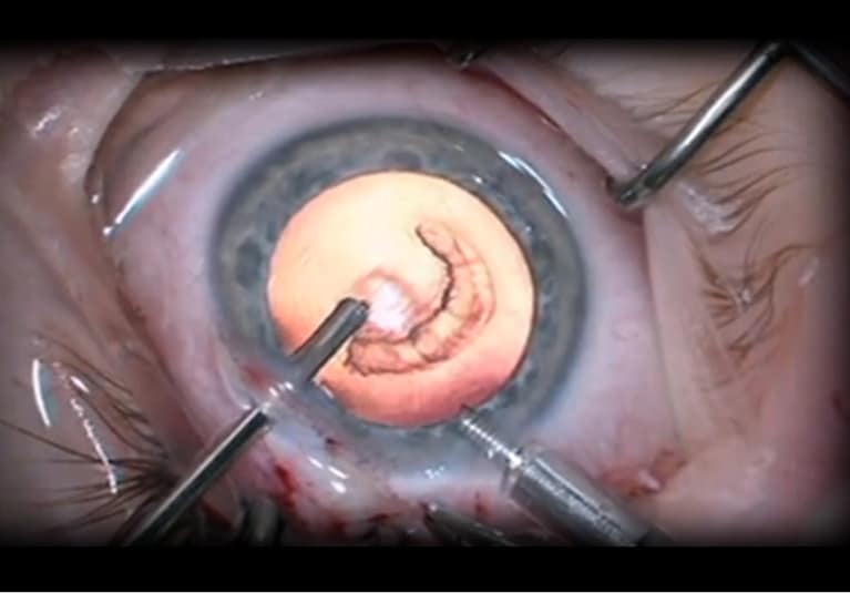
Primary insertion of an intraocular lens during pediatric cataract surgery is the standard of care for children age 2 years and older. Although intraocular lens implantation has become more common in patients younger than 2 years, concerns about adverse events, re-operations and refractive changes continue to be serious considerations in infants.

To evaluate these concerns, Erick D. Bothun, M.D., and a research team with Ophthalmology at Mayo Clinic in Rochester, Minnesota, conducted a retrospective review of infants ages 7 to 24 months treated by surgeons at 10 Infant Aphakia Treatment Study (IATS) sites during that study’s enrollment period.

“The IATS assessed the outcomes and complications of unilateral cataract surgery in infants ages 1 to 7 months,” says Dr. Bothun. “This study, the Toddler Aphakia and Pseudophakia Study (TAPS), is a retrospective consecutive case series of cataract surgery procedures performed in infants ages 7 to 24 months by surgeons who simultaneously were enrolling younger babies in the IATS.

“Because the surgical and clinical care of the TAPS mirrored that of the IATS, the outcomes of surgery for children ages 7 to 24 months in the TAPS can be compared with those reported for children ages 1 to 7 months in IATS.”

The IATS five-year results from the Infant Aphakia Treatment Study Group appeared in JAMA Ophthalmology in 2014. The first TAPS cohort results were published in Ophthalmology in 2019.

Infants were eligible for inclusion in the TAPS registry if they had undergone unilateral or bilateral cataract surgery performed by an IATS surgeon prior to age 24 months during the IATS enrollment period (Jan. 1, 2004, through Dec. 31, 2010). This first manuscript from the TAPS registry involved only the infants with unilateral cataract, and for this effort, the TAPS exclusion criteria largely paralleled the IATS criteria.

The 10 IATS sites registered 96 infants ages 7 to 24 months with a history of unilateral cataract surgery between 2004 and 2010. Ultimately, 56 infants were included in the unilateral TAPS.

Surgery was performed on the right eye in 31 infants (55%) and on the left eye in 25 infants (45%). A primary intraocular lens was inserted in 51 infants (91%). Intraocular lenses were implanted in 20 of 24 infants (83%) who were 7 to 12 months of age, and in 31 of 32 infants (97%) who were 13 to 24 months of age.

Clinical and surgical records were reviewed for visual acuity, refractive correction, patching compliance, intraocular pressure, ocular motility, and anterior segment and ocular fundus examination findings until the final study visit, when the infants were between 4 and 6 years of age. Other patient details included gender, age at surgery, cataract description, strabismus measurements and intraocular lens power.

Main outcome measures

Intraoperative complications and adverse events were recorded using the IATS criteria. Intraoperative complications occurred in four infants (7%). An additional unplanned intraocular surgery occurred in 14% of infants. Adverse events were identified in 24%, with a 4% incidence of glaucoma suspect. Strabismus surgery was performed in 40% of the infants with strabismus before 4 years of age.

Visual acuity, strabismus, stereopsis and glaucoma outcomes were not statistically different between the study groups. Neither adverse events nor intraocular re-operations were more common for infants with surgery at 7 to 12 months of age than for those who underwent surgery at 13 to 24 months of age.

“Although most infants in TAPS between 7 months and 2 years of age underwent intraocular lens implantation concurrent with unilateral cataract removal, the incidences of complications, re-operations and glaucoma appear much lower than when intraocular lenses were used by the same surgeons in infants younger than 7 months of age in the IATS,” says Dr. Bothun. For infants who received an intraocular lens in IATS, intraoperative complications occurred in 28%, adverse events in 81%, and additional intraocular surgeries in 72%.

“Due to inflammatory risks, structural challenges and characteristics of eye growth, the IATS and other efforts showed that cataract surgery risks increase in infancy compared with older children and teenage youth. Prior to the TAPS, the literature has lacked documentation regarding cataract surgery outcomes in infants just older than the IATS group. The TAPS findings support the relatively safe use of intraocular lenses in infants between 7 and 24 months of age,” says Dr. Bothun.

For more information

The Infant Aphakia Treatment Study Group. Comparison of contact lens and intraocular lens correction of monocular aphakia during infancy: A randomized clinical trial of HOTV optotype acuity at age 4.5 years and clinical findings at age 5 years. JAMA Ophthalmology. 2014;132:676.

Bothun ED, et al. Outcomes of unilateral cataracts in infants and toddlers 7 to 24 months of age: Toddler Aphakia and Pseudophakia Study (TAPS). Ophthalmology. 2019;126:1189.

Posted in Uncategorized | Leave a comment

Nation’s Leading Medical Specialty Organizations Applaud Congressional Leaders’ Introduction of Legislation to Improve Oversight and Transparency in Prior Authorization

WASHINGTON, June 5, 2019 — A coalition of eight national medical specialties today pledged support for the bipartisan Improving Seniors’ Timely Access to Care Act of 2019 (H.R. 3107), legislation that would protect patients from unreasonable Medicare Advantage plan requirements that needlessly delay or deny access to medically necessary care. The legislation is sponsored by Reps. Suzan Delbene, D-Wash., Mike Kelly, R-Pa., Roger Marshall, MD, R-Kan., and Ami Bera, MD, D-Calif. 

Currently, Medicare Advantage plans require physicians to obtain advance approval before physicians can provide certain services to their patients. This prior authorization process is intended to control costs by reducing medically unnecessary tests and procedures. Rather than accomplish this goal, many health plans are now widely using prior authorization indiscriminately, creating hurdles and hassles for patients and their physicians that lead to treatment delays that may endanger their health. 

The process for obtaining this approval is lengthy, typically requiring physicians or their staff to spend the equivalent of two or more days each week negotiating with insurance companies. This time would better be spent taking care of patients, especially because the vast majority of these requests are ultimately approved. 

Physicians report devastating results from the worst prior authorization delays, including blindness, loss of function and tumor growth. 

The Improving Seniors’ Timely Access to Care Act is based on a consensus statementon prior authorization adopted by leading national organizations representing physicians, hospitals and health plans. The bill would improve the current prior authorization system by requiring the Centers for Medicare & Medicaid Services (CMS) to regulate Medicare Advantage plans on prior authorization’s use. 

The legislation would also bring greater transparency to the prior authorization process by requiring Medicare Advantage plans to report to CMS on the extent of their use of prior authorization and the rate of approvals or denials by service and/or prescription medication. Over 100 members of Congress called for such reform in a bipartisan letter to CMS last year. 

In conjunction with the bill’s introduction, the Regulatory Relief Coalition, the group of eight medical specialty societies, released results of a physician survey that details the extent to which abusive prior authorization policies are putting patients at risk and increasing burdens on physician practices.  

The survey results include the following:

  • An overwhelming number of physicians (87%) report that prior authorization has a significant (40%) or somewhat negative (47%) impact on patients’ clinical outcomes.
  • A third of physicians (32%) report that their patients often abandon their treatment because of prior authorization.
  • Three-quarters of physicians (74%) report that during the past five years, stable patients have been asked to switch medications by their health plan even though there was no medical reason to do so.
  • Eighty four percent of physicians report that the burden associated with prior authorization has significantly increased over the past five years, with more than half of all practices subjected to 11 or more requests each week, with many finding these requests exceed 40 per week. 

“For more than two years, the Regulatory Relief Coalition made it our responsibility to chip away at barriers that deny our patients timely access to medically necessary care, and the walls enabling prior authorization’s abuse are about to come down,” said George A. Williams, MD, president of the American Academy of Ophthalmology, one of the coalition’s members.   

Echoing his sentiments, Ann R. Stroink, MD, chair of the American Association of Neurological Surgeons and Congress of Neurological Surgeons Washington Committee, concluded, “With the Improving Seniors’ Timely Access to Care Act, a strong, bipartisan group of elected officials in Congress is saying enough is enough, care delayed is care denied, and America’s seniors deserve the care that they expect from the Medicare program.” 

About the Regulatory Relief Coalition

The Regulatory Relief Coalition is a group of eight national physician specialty organizations advocating for a reduction in Medicare program regulatory burdens to protect patients’ timely access to care and allow physicians to spend more time with their patients. 

 American Academy of Neurology ¨ American Academy of Ophthalmology ¨ American Association of Neurological Surgeons ¨ American College of Cardiology ¨ American College of Rheumatology ¨ American College of Surgeons ¨ American Urological Association ¨ Congress of Neurological Surgeons

Posted in Uncategorized | Leave a comment